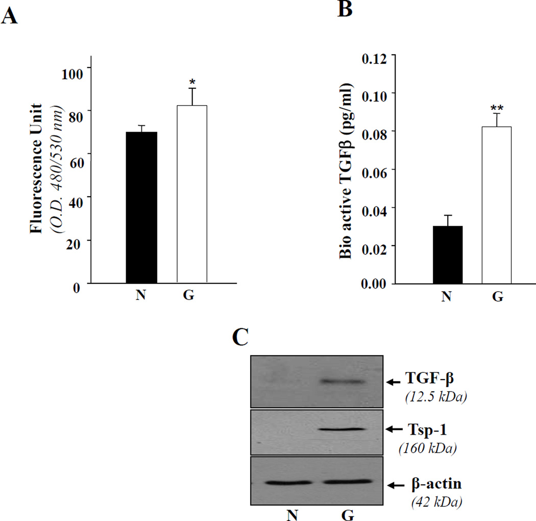
Figure 2. TM cells derived from glaucomatous subjects showing increased expression of ROS.
A, cells were cultured in 96 well plates containing Opti-MEM plus 10% FBS as described in Materials and Methods. The next day, the medium was replaced with HBSS containing 5–10µg of H2-DCFH-DA, and fluorescence intensity was measured using DTX 880, Multimode detector, adjusted at 485nm (excitation) and 535nm (emission) (Beckman Coulter). Histogram values are means ± SD of 3 independent experiments, each with triplicate wells, showing increased levels of ROS in glaucomatous TM cells (G, open bar, *p< 0.05). B, Glaucomatous TM cells secrete bioactive TGFβ. Cells were cultured in 96 well plate for 72 h, culture supernatant was collected, and bioactive TGFβ was assessed using Emax immunoassay system (Promega). Equal numbers of nonglaucomatous cells were also cultured and supernatant was used as control. A higher level of TGFβ1 was detected in glaucomatous cells (B, open bar, **p< 0.001) in comparison to nonglaucomatous TM cells. From another set of experiments, cells extract was prepared and used for Western analysis. The level of TGFβ1 protein was comparatively higher in glaucomatous TM cells (C, upper panel). To see the expression level of Tsp-1, a protein implicated in activation of TGFβ and glaucoma pathogenesis, the membrane stained earlier with PRDX6 or β-actin antibody, was stripped and reprobed with Tsp-1 antibody. The results revealed an increased level of Tsp-1 (C, right lane; G).

