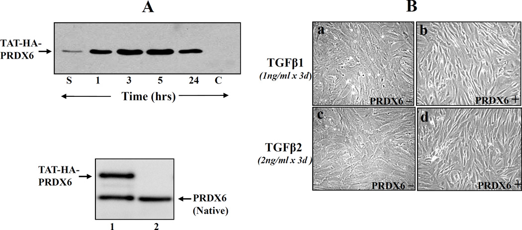
Figure 4. Western analysis showing internalization of TAT-HA-PRDX6 in TM cells.
A, Recombinant PRDX6 linked TAT were generated and purified as described earlier [7, 8]. The protein was supplied to the culture medium at a concentration of 4 µg/ml and transduction efficiency of TAT-HA-PRDX6 was evaluated at 1, 3, 5, and 24h. HA-PRDX6 was used as control (lane, c). 3 h later, cells were washed several times, cell extracts were prepared, and Western blot was carried out with anti-His antibody (Invitrogen). Lane S, culture medium after 1 h of addition; Protein band corresponding to lanes 1, 3, 5, 24 denotes time after TAT-HA-PRDX6 protein; lane C, control (HA-PRDX6 without TAT). Results showed that TAT-HA-PRDX6 internalized into cells while HA-PRDX6 did not. A, lower panel showed protein bands of TAT-HA-PRDX6 (lane1 upper band) and native PRDX6 (lane 1 and 2, lower band). To visualize delivered PRDX6 and native PRDX6, membrane was probed with PRDX6 monoclonal antibody and visualized as described in ‘Materials and Methods’ section. (B) Photomicrographs showing that a supply of PRDX6 attenuated TGFβ1 and TGFβ 2 induced adverse changes in TM cells (passage (P3), and restored normal phenotypes. TM cells derived from nonglaucomatous subjects (P3) were cultured in Opti-MEM plus 10% FBS. These cells were supplied with TAT-HA-PRDX6 or every third day up to P12. Photomicrographs were taken at P8 and are representative of three experiments.

