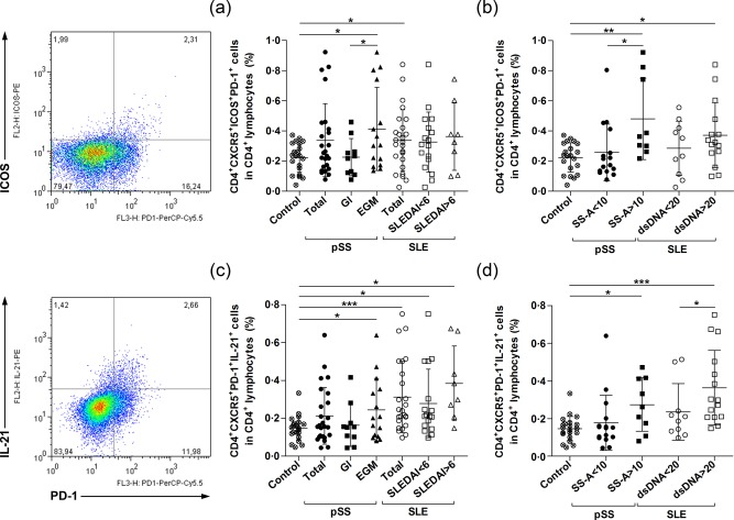
Figure 2.

Enumeration of circulating follicular T helper cells (Tfh)‐like cells in primary Sjögren's syndrome (pSS) patients, systemic lupus erythematosus (SLE) patients and healthy subjects. Peripheral blood mononuclear cells (PBMCs) were isolated from 25 pSS patients; 25 SLE patients and 21 healthy controls were then stained with labelled antibodies as described previously. Circulating Tfh‐like cells were assessed as their percentage in the CD4+ lymphocyte population. Representative dot‐plots are shown the frequency of ICOS+programmed death 1 (PD‐1)+ and PD‐1+interleukin (IL)‐21+ cells within the CD4+CXCR5+ cell population. (a) Percentages of CD4+CXCR5+ICOS+PD‐1+ Tfh‐like cells in the overall pSS patient population (n = 25), pSS with only glandular symptoms (n = 10), pSS with extraglandular manifestations (EGMs) (n = 15), total SLE patient group (n = 25), SLE with SLE Disease Activity Index (SLEDAI) < 6 (n = 17), SLE with SLEDAI > 6 (n = 8) and control subjects (n = 21). (b) Ratio of CD4+CXCR5+ICOS+PD‐1+ Tfh‐like cells according to the presence of autoantibodies; in anti‐Ro/SSA antibody‐negative group (n = 16), anti‐Ro/SSA antibody‐positive group (n = 9), anti‐dsDNA negative group (n = 10), anti‐dsDNA‐positive group (n = 15) and control subjects (n = 21). (c) Distribution of IL‐21‐producing CD4+CXCR5+ PD‐1+ Tfh‐like cells. (d) Percentages of IL‐21‐producing CD4+CXCR5+PD‐1+ Tfh‐like cells according to the presence of autoantibodies in patient groups. Each data point represents an individual subject; horizontal lines show the mean values with standard deviation (s.d.). Statistically significant differences are indicated by *P < 0·05; **P < 0·01; ***P < 0·001.
