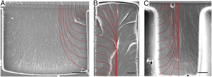
Fig. 4.
(A–C) SEM images show the fracture structure of LCP after polymerization in a 1D microchannel (A), in a pore (B), and between two pillars (C). Escaping director field of LC can be observed from the orientation of the fracture structures in all three structures. The red dotted lines in A–C represent local director field of LCM mirrored in the other half of images. (Scale bars: A, 5 µm; B and C, 2 µm.)

