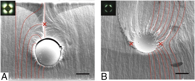
Fig. 5.
(A and B) SEM images of silica colloids suspended in LCP where either a point defect (A) or a line defect (B) was formed to screen the charge of the colloid. (A and B, Insets) POM images of point (A) and line (B) defects circumscribing silica colloids. The director field of the LC is represented by the red dotted line and the red crosses show the positions of defects. (Scale bars: 2 µm.)

