Fig. 4.
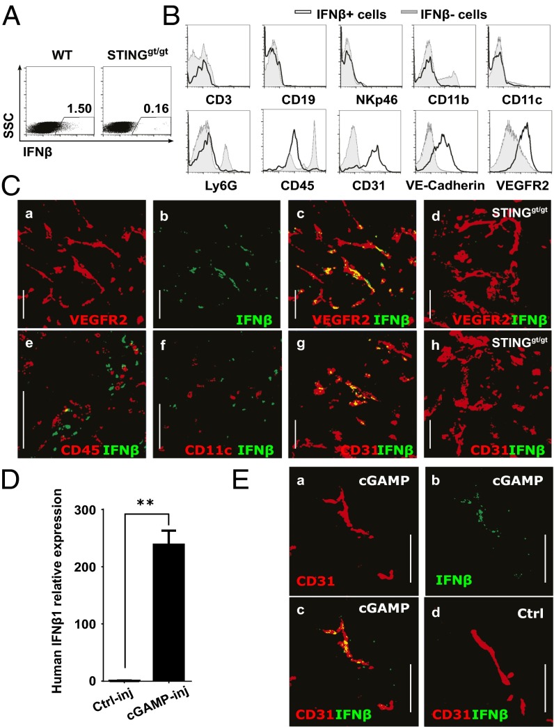
Endothelial cells represent the main IFN-β producers upon intratumoral cGAMP injection in mouse and human. (A–C) WT or STINGgt/gt mice bearing 5-d established s.c. B16F10 tumors were treated by i.t. injection of cGAMP plus Brefeldin A. Tumors were harvested after 5 h for flow cytometry and confocal microscopy analysis. (A) Intracellular IFN-β detection in tumor single cell suspensions. (B) Expression of CD3, CD19, NKp46, CD11b, CD11c, Ly6G, CD45, CD31, VE-cadherin, and VEGFR2 in IFN-β–producing cells (black line, gated as shown in A compared with nonIFN-β–producing cells (gray line). Data are representative of at least three independent experiments. (C) IFN-β expression on CD31 or VEGFR2 cells in tumor cryosections derived from WT (a–c and e–g) or STINGgt/gt mice (d and h). (Scale bar, 100 µm.) (D and E) Resected human melanoma skin metastases were injected with 2′3′-cGAMP (cGAMP-inj) or Lipofectamine alone (Ctrl-inj) and cultured in the presence of Brefeldin A (only in E) for 5 h before analysis. Depicted are: (D) Quantitative real-time PCR analysis of IFN-β1 mRNA expression in treated tumor explants. **P < 0.01 by unpaired t test. (E) Confocal microscopy of IFN-β and CD31 stained on cryosections derived from treated tumor explants. (Scale bar, 100 µm.)

