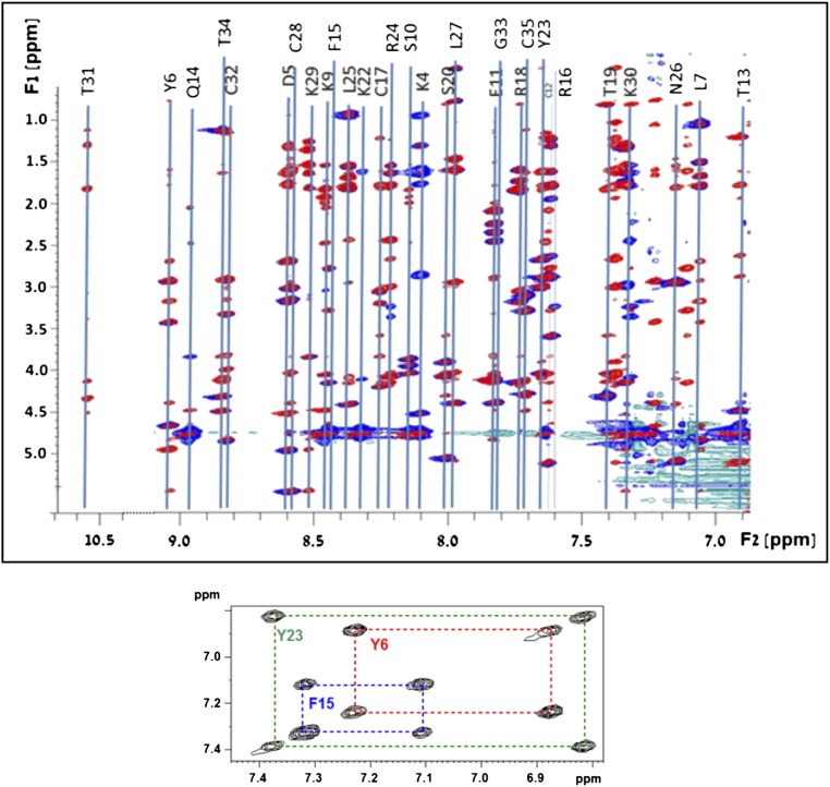
Fig. S3.
NMR determination of the Hui1 structure with method details. Homonuclear fingerprint spectra of Hui1. (Top) HN{F2}/Hali{F1} overlaid regions of the 2D TOCSY (red) and NOESY (blue) spectra of Hui1 acquired in H2O-based buffer. Line connecting spin systems and assignments are shown. (Bottom) Haro{F2}/Haro{F1} region of the TOCSY spectrum with assignments of the three aromatic spin systems.

