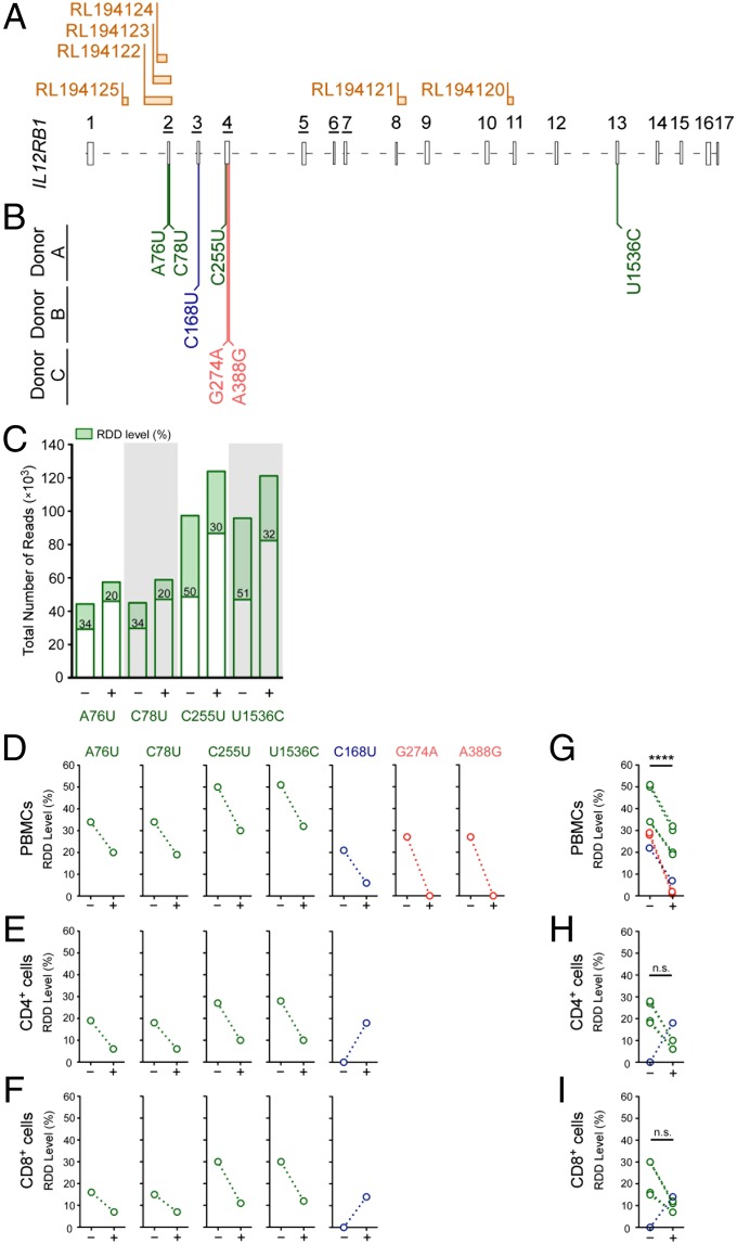
Fig. 1.
IL12RB1 mRNAs contain multiple RDDs, the appearance of which decrease with activation. (A) IL12RB1 mRNA comprises 17 exons, the relative sizes and orientation of which are depicted 5′-3′ alongside the location of six putative R-loop-forming regions: RL194, RL194122, RL194123, RL194124, RL194121, and RL194120. The exon numbers that are underlined (exons 2–7) encode the IL12Rβ1 CBR. (B) The position and nature of the IL12RB1 RDDs in PBMCs of three healthy, immunocompetent donors (donors A, B, and C), as identified by next-generation sequencing. In the top row are the IL12RB1 RDDs present in donor A (A76U, C78U, C255U, and U1536C); the center and bottom rows, respectively, list the IL12RB1 RDDs present in donor B (C168U) and donor C (G274A, A388G). (C) Sequencing read coverage data and relative appearance of each donor A RDD in the absence (−) or presence (+) of an activation signal (PHA); each donor A RDD is listed along the x-axis. Each bar represents the total number of reads at that specific site in the IL12RB1 transcript; the RDD level is indicated in the green portion of each bar. (D) The levels of each individual IL12RB1 RDD found in donors A–C PBMCs in the presence or absence of activation. Each open circle represents one of the RDDs listed in B, using the same color scheme for each donor. An identical analysis is shown for (E) CD4+ and (F) CD8+ cells from the same donor PBMC preparations. (G–I) IL12RB1 RDD-level data were combined from donors A–C to show (G) cumulative RDD levels in donor A–C PBMCs, (H) cumulative RDD levels in donors A and B CD4+ cells, and (I) cumulative RDD levels in donor A and B CD8+ cells, both in the presence and absence of PHA. For A and B, exon boundaries and ribonucleotide number assignments, we used the IL12RB1 ribonucleotide numbering system described in Dataset S6. R-loop sites were identified by Wongsurawat et al. (8); these sites and their designations are taken from their publically available R-loop database found at rloop.bii.a-star.edu.sg/. For statistical comparisons of PHA− and PHA+ data in G and H, donor data were pooled and significance (P) determined using a paired t-test. ****P < 0.0001.

