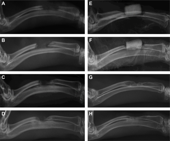
Figure 7.
Imaging examination at day 1 and 4, 8, and 12 weeks.
Notes: (A–D) In the control group, no new bone formation, and the bone defect was not restored. With the extension of time, the broken end gradually hardened. At 12 weeks, the broken ends sclerosed and closed. (E–H) In the experimental group, the material gradually degraded and was absorbed, and there was new bone formation. At 12 weeks, much of the material had degraded and was absorbed, and was only faintly visible with bone defect recovery.

