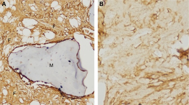
Figure 9.
At 12 weeks after treatment, immunohistochemical examination of collagen I fibers at the lesion (×200).
Notes: (A) Implanting group: a great deal of collagen fibers formed around the material. (B) Control group: collagen fiber formation was not notable.
Abbreviations: F, collagen I fiber; M, material.

