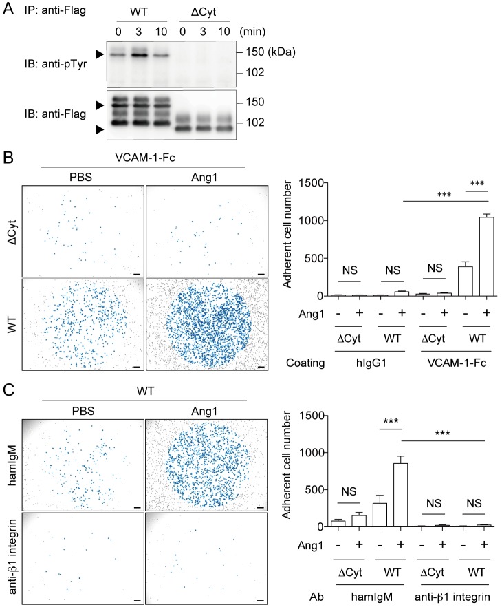
Fig 3. Tie2 signaling enhances MEDMC-BRC6 adhesion to VCAM-1 through α4β1 integrin.
(A) WT/MEDMC-BRC6 and ΔCyt/MEDMC-BRC6 transfectants were established as described in the Methods. Transfectants were stimulated with Ang1 (250 ng/mL), and immunoprecipitated (IP) with an anti-Flag mAb. Tyrosine phosphorylation of Tie2 was analyzed by blotting with anti-pTyr Ab. (B) WT/MEDMC-BRC6 and ΔCyt/MEDMC-BRC6 were treated with Ang1 (250 ng/mL) or not and incubated on hIgG1- or VCAM-1-Fc-coated wells. Adherent cells, as observed blue-colored, were counted by using a microscope (20 mm2 per well). (C) Neutralizing anti-β1 integrin Ab (20 μg/mL) or control Ab (20 μg/mL) was added under the conditions described in B. Cells were incubated on VCAM-1-Fc-coated wells, and adherent cells were similarly counted. WT, WT/MEDMC-BRC6; ΔCyt, ΔCyt/MEDMC-BRC6. Scale bars, 200 μm. Data show mean values ± SEM (n = 3 or 5). ***p < 0.001.

