Figure 1.

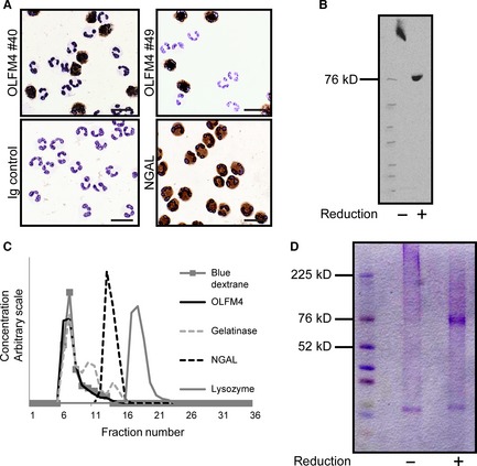
Identification of OLFM4 in Neutrophils. (A) Cytospins of neutrophils isolated from peripheral blood were immune‐stained using OLFM4 antibodies from clone #40 and #49. Clearly only a fraction of neutrophils stain positive for OLFM4 whereas all neutrophils stain positive for NGAL. (B) Western blotting of isolated specific granules using anti‐OLFM4 antibody #49 under reducing and non‐reducing conditions. (C) Size exclusion chromatography of OLFM4 from specific granules of human neutrophils. Distribution of markers was determined by ELISA or by absorption of light (Blue Dextran). (D) Coomassie blue stained gels of affinity purified OLFM4 under reducing and non‐reducing conditions.
