Figure 2.

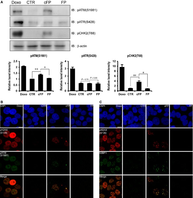
Activation of DNA damage response in cFP‐treated INT‐407 cells. (A) CHK2 (T68) and ATM (S1981) are phosphorylated in cFP‐treated INT‐407 cells. After cells were incubated with 1 mM cFP or linear FP dipeptides (FP) for 48 hrs, the lysates were immunoblotted with anti‐phospho ATR (S428), anti‐phospho ATM (S1981) and anti‐phospho CHK2 (T68) antibodies respectively. As controls, INT‐407 cells were untreated (CTR) or treated with 1 μM doxorubicin (Doxo). Western blots were analysed quantitatively. (B and C) Representative images demonstrate the co‐localization of phosphorylated H2AX (S139), phosphorylated ATM (S1981) and 53BP1 in cFP‐treated INT‐407 cells. The cells treated with 1 mM cFP or linear FP dipeptides (FP) for 48 hrs were co‐immunostained with anti‐phospho H2AX (S139), anti‐phospho ATM (S1981) and anti‐53BP1 antibodies in combination. As controls, INT‐407 cells were untreated (CTR) or treated with 1 μM doxorubicin (Doxo); scale bar, 10 μm. *P ≤ 0.05 and **P ≤ 0.01.
