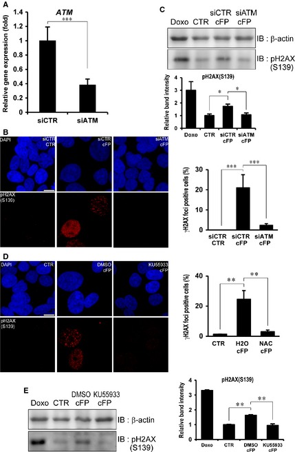
Figure 3.

ATM‐mediated DNA damage in cFP‐treated INT‐407 cells. (A) The transcript of ATM was efficiently depleted by ATM siRNA. The expression of ATM was determined by real‐time PCR in control (siCTR) or ATM siRNA (siATM). (B) Representative images demonstrate that depletion of ATM decreases γH2AX foci formation in cFP‐treated INT‐407 cells. After control or ATM siRNA transfection, cells were incubated with 1 mM cFP for 48 hrs and immunostained with an anti‐phospho H2AX (S139) antibody. Untreated cells (CTR) were used as a negative control; scale bar, 10 μm. (C) Depletion of ATM decreases phosphorylation of H2AX (S139) in cFP‐treated INT‐407 cells. After siRNA transfection, cells were incubated with 1 mM cFP for 48 hrs and lysates were immunoblotted with anti‐ phospho H2AX (S139) antibody. As controls, INT‐407 cells were untreated (CTR) or treated with 1 μM doxorubicin (Doxo). Western blots were analysed quantitatively. (D) Representative images demonstrate that enzyme activity of ATM is required for cFP‐induced γH2AX foci formation. After cells were incubated with 1 mM cFP in conjunction with 10 μM ATM inhibitor (KU‐55933) or DMSO for 48 hrs, cells were immunostained with anti‐ phospho H2AX (S139) antibody. Untreated cells (CTR) were used as a negative control; scale bar, 10 μm. (E) After cells were incubated with 1 mM cFP in conjunction with 10 μM ATM inhibitor (KU‐55933) or DMSO for 48 hrs, the lysates were immunoblotted with anti‐ phospho H2AX (S139) antibody. As controls, INT‐407 cells were untreated (CTR) or treated with 1 μM doxorubicin (Doxo). Western blots were analysed quantitatively. *P ≤ 0.05, **P ≤ 0.01 and ***P ≤ 0.005.
