
Figure 1.
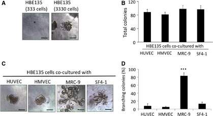
Effects of mesenchymal cells on colony formation in 3D culture. (A) Phase‐contrast images of HBE135 cells cultured alone in rBM. HBE135 cells did not form colonies at a low cell density (333 cells in 100 μl rBM, left panel), but formed spherical colonies at a high cell density (3330 cells in 100 μl rBM, right panel). (B–D) HBE135 cells were cultured in rBM with various types of mesenchymal cells; scale bars 100 μm. (B) Numbers of colonies formed by HBE135 cells in coculture with mesenchymal cells (3 × 10³ cells/ml) in rBM. Each value represents the mean ± S.D. (n = 4). (C) Phase‐contrast images of HBE135 cell colonies co‐cultured with mesenchymal cells [human umbilical vein endothelial cells (HUVECs), human microvascular endothelial cells (HMVECs), MRC‐9 cells and SF4‐1 cells]; scale bars 100 μm. (D) Numbers of colonies formed by of HBE135 cells with a complex branching structure. Each value represents the mean ± S.D. (n = 4); scale bars: 100 μm. ***P < 0.001