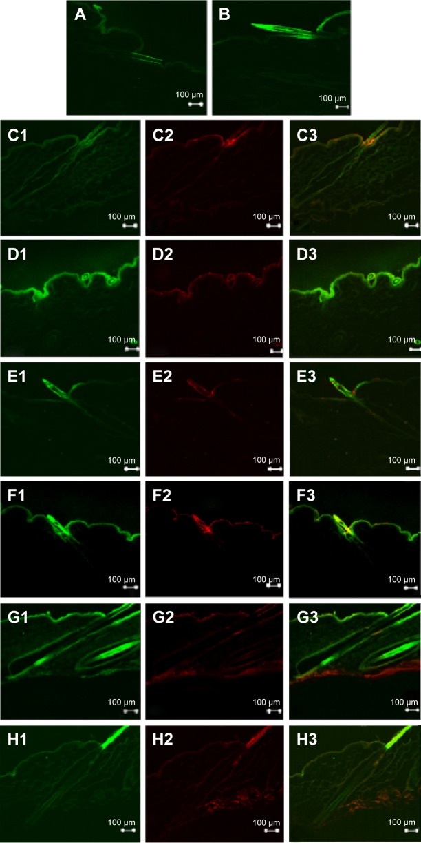
Figure 4.
Confocal images of the skin cross section obtained at 4 hours after deposition of (A) NaFI solution without SN, (B) NaFI solution with SN, (C) NaFI-loaded-Rh-PE-labeled liposomes: CL without SN, (D) NaFI-loaded-Rh-PE-labeled liposomes: CL NaFI-loaded-Rh-PE-labeled liposomes: CL with SN, (E) NaFI-loaded-Rh-PE-labeled liposomes: PL without SN, (F) NaFI-loaded-Rh-PE-labeled liposomes: PL with SN, (G) NaFI-loaded-Rh-PE-labeled liposomes: PL-LI without SN, and (H) NaFI-loaded-Rh-PE-labeled liposomes: PL-LI with SN.
Notes: In images C to H, it is divided into 3 parts: 1) green fluorescence of NaFl; 2) red fluorescence of Rh-PE; and 3) overlay of green fluorescence of NaFl and red fluorescence of Rh-PE. The scale bar represents 100 µm. All confocal images were obtained at a magnification of ×10.
Abbreviations: CL, conventional liposome; NaFI, sodium fluorescein; PL, PEGylated liposome; PL-LI, PEGylated liposome with d-limonene; Rh-PE, rhodamine B 1,2-dihexadecanoyl-sn-glycero-3-phosphoethanolamine triethylammonium salt; SN, sonophoresis.

