Figure 3.
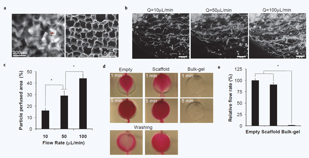
Characterization of inter-scaffold hydrodynamic microenvironment. (a) Fluorescent image of micro-scale pores in a scaffold-chip visualized by injecting rhodamine solution (Left) and SEM image of subcellular scale pores in bulk hydrogel matrix (Right). (b) Perfusion profile of cellular size fluorescent microsparticles (D = 10∼15 µm) at three different flow rates. Streamline flows were captured by setting exposure time to 20 seconds. (Scale bar, 200 µm) (c) Quantitative comparision of perfused area at different flow rates. Increased flow rates perfuse not only more particles but also more area. (n = 5, *p < 0.05) (d) Perfusion profiles of rhodamine solution in an empty, scaffold and bulk-gel inserted chip under gravity-induced flow. No significant mass transport was detected in a bulk gel inserted chamber due to high fluidic resistance in the absence of macroscopic pores. Considerably longer retention of rhodamine molecule was observed in a scaffold-chip than an empty chamber. (e) Comparison of relative flow rates. Regularly arranged, hierarchical scale pores in hydrogel scaffold supports about 90% fluidic perfusion of an empty chamber (n = 5, *p < 0.1).

