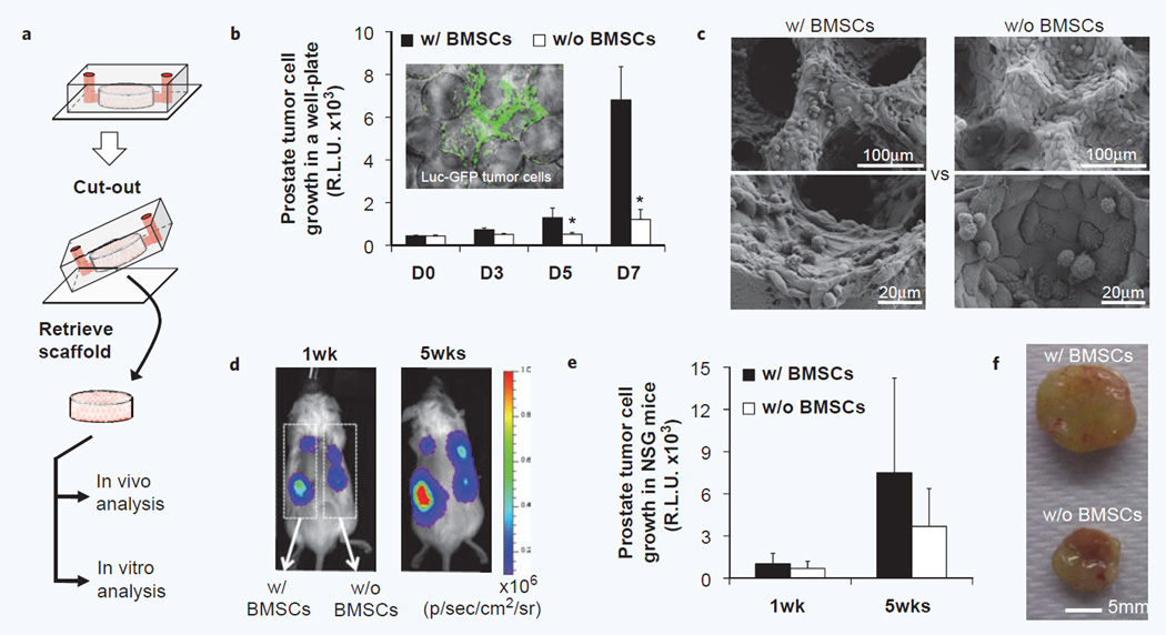
Figure 5.
Retrieval of hydrogel scaffold from microfluidic devices with retained xenograft capacity. (a) Schematic of retrieval of an integrated hydrogel scaffold from a microfluidic device for continues in vitro and in vivo analysis. (b) In vitro growth analysis of microfluidically captured Luc-GFP PC3 prostate tumor cells after transferring to a well plate using bioluminescent imaging (n = 6, *p < 0.05). Fluorescent image of tumor cells in a retrieved scaffold (Inner panel). (c) Morphological analysis of tumor cell growth with and without human BMSCs after 3 weeks in vitro culture. (d) In vivo growth analysis after subdermal implantation into immunodeficient mice using bioluminescent imaging. (e) Quantification of bioluminescent monitoring of tumor cell growth over 5 weeks in vivo (n = 6). (f) Gross image of explanted scaffolds 7 weeks after implantation.

