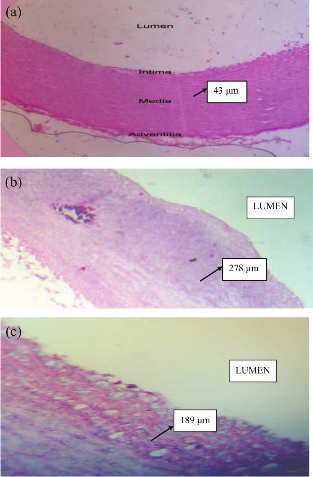
Figure 1.

The photomicrograph of histomorphometric section in aortic arch of (a) normal, (b) hyperlipidemic, and (c) sitagliptin hyperlipidemic rabbits shows the difference in aorta intima-media thickness. The section stained with hematoxylin and eosin (4×): (a) image showing normal appearance of arterial wall layers; (b) image showing a fibro-atheromatous plaque with thick layers of fibrous connective tissue overlying a largely necrotic, fatty mass—advance atherosclerotic lesion (Type IV); and (c) image showing significant decrease in the aortic intima thickness as compared to induced untreated (b) group.
