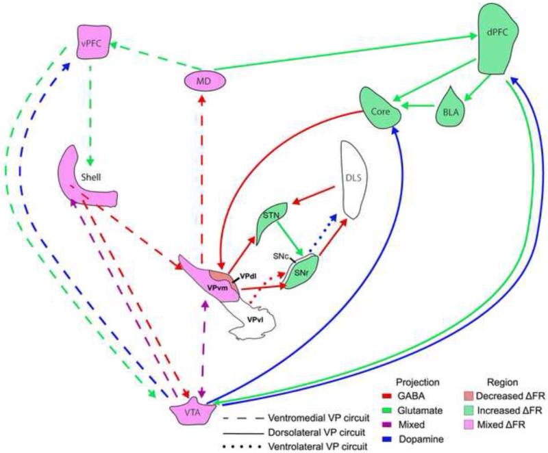
Figure 7. Proposed changes in firing rate during responses for drugs of abuse involving the VP subregions.
Neurons of the AcbC exhibit increased firing rates during responses for cocaine (Ghitza et al. 2004), which are likely the result of dPFC and BLA activation during the same behavior (Chang et al., 1997, 2000; Carelli et al., 2003). Activation of AcbC leads to decreased firing rates of VPdl neurons during the response (Root et al., 2013) and likely disinhibits its target structures SNr and STN (Lardeux et al., 2013; Fan et al., 2012). While AcbSh neurons are less sensitive to the response compared with AcbC neurons, AcbSh neurons exhibiting changes in firing rate during the response are heterogeneous (Ghitza et al., 2004). Such heterogeneity is found within the VPvm subregion (Root et al., 2013) and is likely to continue within its target regions VTA and MD. The involvement of VPvl and its circuits (SNc and DLS; dotted lines with arrows) is unclear (white color). Dashed lines with arrows represent circuits involving VPvm. Solid lines with arrows represent circuits involving VPdl. Purple shaded structure indicates heterogeneous (increased or decreased) change in firing rate during cocaine self-administering responses. Red shaded structure and green shaded structure indicates decreased and increases change in firing rate during cocaine self-administering responses. The illustrated subregions are proposed to represent subregions throughout the anteroposterior extent of the VP.

