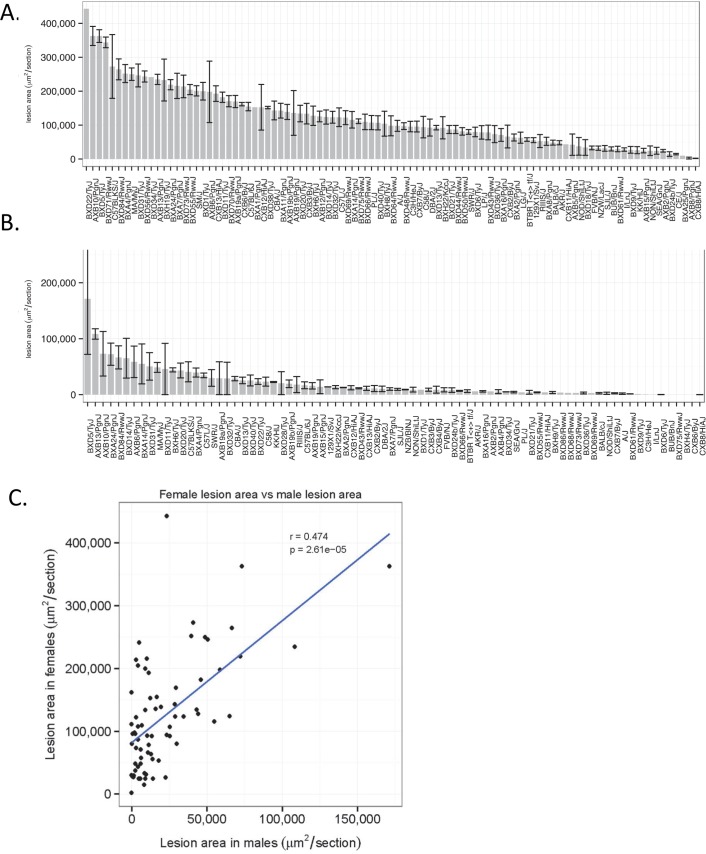
Fig 4. Atherosclerosis in Ath-HMDP mice.
Atherosclerotic lesion size (μm2 ± SEM) in the proximal aorta and aortic sinus were quantitated for 697 female mice (A) and 281 male mice (B) using oil red—O staining. In each panel, strains are arranged in rank order by strain-average lesion area. (C) Correlation between strain-average lesion areas in male and female Ath-HMDP mice.

