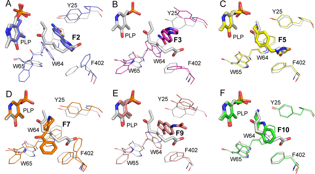
Figure 2.
Fragment binding induces side chain conformational differences. The KAPA-bound reference structure (gray) is compared to a different structure in each panel: (A) F2; (B) F3; (C) F5; (D) F7; (E) F9; (F) F10. The common orientation of view of all complexes underscores the differences in the position and orientation seen in the binding of different fragments.

