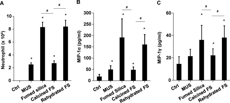
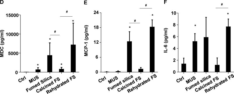
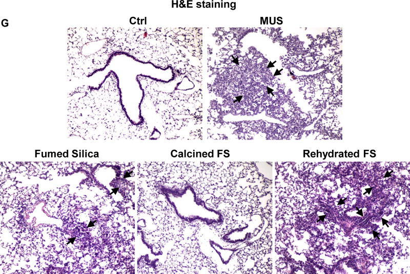
Figure 2. The acute pro-inflammatory effects of fumed silica in the lung is attenuated by calcination but exacerbated by rehydration.
C57BL/6 (n=6) mice were exposed to 1.6 mg/kg of pristine, calcined and rehydrated fumed silica nanoparticles by oropharyngeal aspiration for 40 h, then animals were sacrificed to collect BAL fluid and lung tissue. (A) Neutrophil count. *p<0.05 compared to control mice. #p<0.05 compared to calcined fumed silica-treated mice. An ELISA microarray assay was used to measure the following cytokine and chemokine levels: including (B) MIP-1α, (C) MIP-1γ, (D) MDC, (E) MCP-1 and (F) IL-6. MIN-U-SIL (MUS), a natural form of crystalline silica, was used as a positive control. *p<0.05 compared to control mice. #p<0.05 compared to calcined fumed silica-treated mice. (G) H&E staining showing focal inflammation induced by fumed silica. Calcination of the particles attenuated lung inflammation but rehydrated fumed silica nanoparticles exacerbated the inflammatory effects. The regions of focal inflammation are indicated by the arrows.


