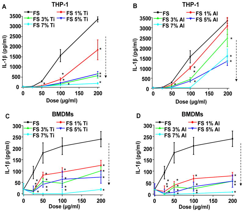
Figure 7. Reduction in IL-1β production by Ti and Al doping of fumed silica nanoparticles.
(A–B) IL-1β production induced by doped fumed silica in THP-1 cells. Naive THP-1 cells were treated with PMA (1 μg/mL) for 16 h. PMA-differentiated THP-1 cells were then exposed to 25–200 μg/mL of (A) Ti-doped and (B) Al-doped fumed silica nanoparticles for 24 h in the presence of LPS (10 ng/mL). IL-1β production was quantified by ELISA. *p<0.05 compared to non-doped fumed silica. (C–D) IL-1β production induced by doped fumed silica in bone marrow-derived macrophages (BMDMs). BMDMs obtained from wild type C57BL/6 mice were exposed to 25–200 μg/ml of (C) Ti-doped and (D) Al-doped fumed silica nanoparticles for 24 h in the presence of LPS (500 ng/mL). *p<0.05 compared to non-doped fumed silica.

