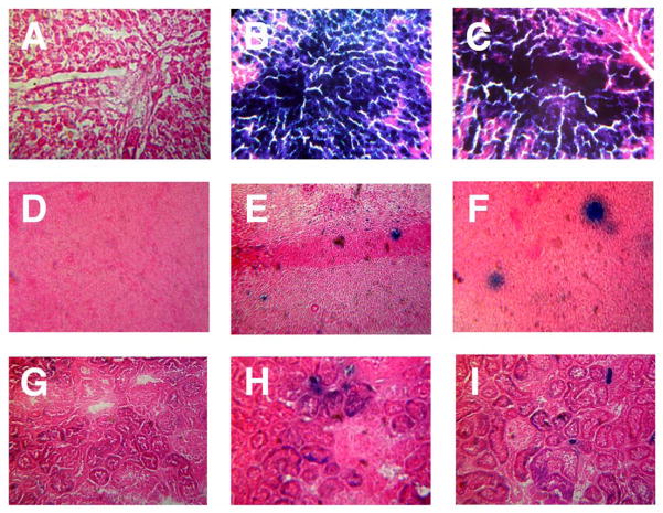
Figure 3. Transgene Expression in Key Tissues Four Days After Administration of a Single Dose of Recombinant Adenovirus by Different Routes.
Male Sprague Dawley rats were given either phosphate buffered saline (vehicle control, Column 1) or 5.7 × 10¹² vp/kg of adenovirus expression beta-galactosidase by injection into the jugular vein through an implanted catheter (Column 2) or by direct injection into lateral tail vein (Column 3). Animals were sacrificed 4 days after treatment and liver (Panels A–C), spleen (Panels D–F) and kidney (Panels G–I) were evaluated for transgene expression by X-gal histochemistry. Morphological assessment was performed from serial sections of three rats for each treatment group and transgene expression assessed by visual inspection of the tissue for the blue product generated by active beta-galactosidase in the presence of the substrate 5-bromo-4-chloro-3-indolyl-β-D-galactoside (X-gal). Sections from animals given saline did not stain positive for the transgene while those from animals dosed via the jugular cannula contained slightly more of the transgene than those dosed by the tail vein. Magnification: Panels A–C, 400 ×, Panels D–F, 300 ×, G–I, 150 ×.

