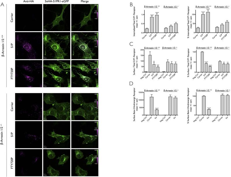
Figure 5. Role of β-arrestins during ligand induced internalization of S1PR1.
(A) MEF cells from mice expressing (β-Arrestin1/2+/+) or not (β-Arrestin1/2−/−) β-arrestin1 and β-arrestin2 and stably expressing 3xHA-S1PR1-eGFP were incubated with anti-HA antibody in the presence of DMSO only (Carrier) or the ligands S1P or FTY720P. Images for each condition correspond to a center plane obtained using spinning disc confocal microscopy. An acid wash step at the end of the experiment was used to remove most of the surface bound anti-HA antibodies. Scale bar 10 μm.
(B) Quantification of the internalization data obtained in panel (A) expressed as mean +/− sem. Normalized data correspond to the values obtained using β-arrestin 1/2 +/+ cells in the presence of S1P. Number of β-arrestin 1/2 +/+ cells analyzed were 27, 37 and 38 for Carrier, S1P, and FTY720P, respectively; number of corresponding β-arrestin 1/2 −/− cells were 37, 33 and 34. The symbol **** highlights that the difference between carrier and ligands were statistically significant with p values of < 0.0001.
(C) Flow cytometry analysis of S1PR1 uptake in the presence (β-arrestin 1/2 +/+) or simultaneous absence (β-arrestin 1/2 −/−) of both β-arrestins. The data is from 5 independent experiments. The symbols ** and *** highlight that the difference between carrier and ligand were statistically significant with p value < 0.0001 and 0.004 respectively. Data on the right panel were normalized to the values obtained using β-arrestin 1/2 +/+ cells in the absence of ligand (Carrier). The symbols **** and ** indicate statistical significance difference with p values of <0.0001 and 0.008 respectively.
(D) Flow cytometry analysis of β2AR uptake in the presence (β-arrestin 1/2 +/+) or simultaneous absence (β-arrestin 1/2 −/−) of both β-arrestins. Cells stably expressing Flag-β2-AR were treated with (Iso) or without (Carrier) 25μM Isoproterenol at 37°C for 25 minutes. The amount of surface receptor was determined by antibody labeling followed by flow cytometry. Data on the right panel normalized to the values obtained using β-arrestin 1/2 +/+ cells in the absence of ligand (Carrier). The data is from 6 independent experiments, 3 of which were conducted simultaneous to those in panel C. The symbol **** highlights that the difference between carrier and ligand were statistically significant with a p value < 0.0001.

