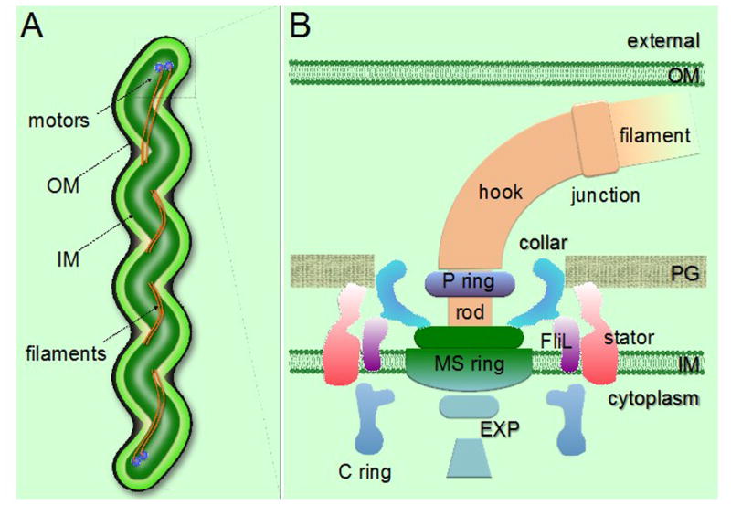
Figure 2.

General morphology and periplasmic flagellar structures in spirochetes. (A) Schematic model of a spirochete cell showing the periplasmic flagellar filaments located between the outer membrane (OM) and the inner membrane (IM), causing the characteristic flat-wave morphology. (B) Schematic model of the periplasmic flagellar motor illustrating various flagellar motor components. PG, peptidoglycan layer; EXP, export apparatus.
