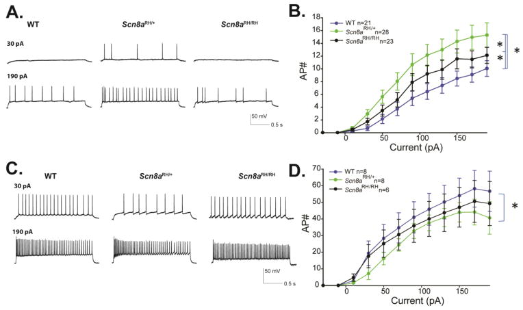
FIGURE 5.
Firing properties of CA3 neurons from R1627H mice. A. Sample traces from CA3 pyramidal neurons of WT, Scn8aRH/+ and Scn8aRH/RH mice at depolarizing current injections of 30 and 190 pA from a resting potential of −70mV. B. Average number of action potentials (AP#) plotted against current injection (pA) (Error bars represent SEM, n = number of cells ; *P < 0.05, two-way ANOVA, Holm-Sidak correction). C. Sample traces from CA3 interneurons of WT, Scn8aRH/+ and Scn8aRH/RH mice at depolarizing current injections of 30 and 190 pA from a resting potential of −70 mV. B. Average number of action potentials (AP#) plotted against current injection (pA). Error bars represent SEM, n = number of cells, *P < 0.05 two-way ANOVA, Holm-Sidak correction.

