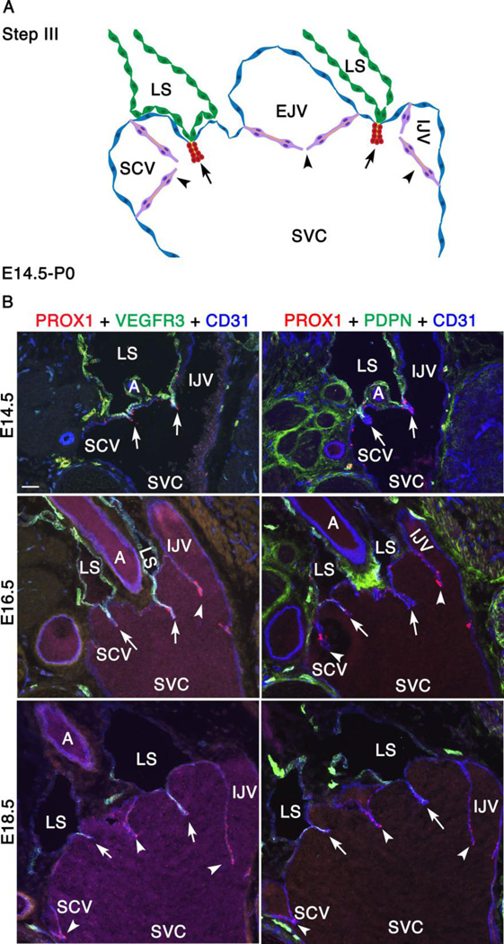
Figure 4. Between E14.5 and P0, VVs are formed and LEC markers are gradually downregulated in lymph sacs.
(A) This is the third and final step of LVV development between E14.5 and P0. The schematic depicts the overall arrangement of the LVV complex in frontal orientation. The only obvious change during this interval is the appearance of VVs at E16.5. VVs are in magenta. Rest of the cells is color coded as in Figure 2. (B) Immunohistochemistry revealed that the LEC markers VEGFR3 and podoplanin (PDPN) are gradually downregulated in LSs. However, they remain strongly expressed in the LECs that form LVVs (arrows).
Statistics: n= 3 for each developmental stage.
Abbreviations: LS, lymph sac; IJV, internal jugular vein; EJV, external jugular vein; SCV, subclavian vein; SVC, superior vena cava.
Scale bars: 50 µm for B and 1 µm for C.

