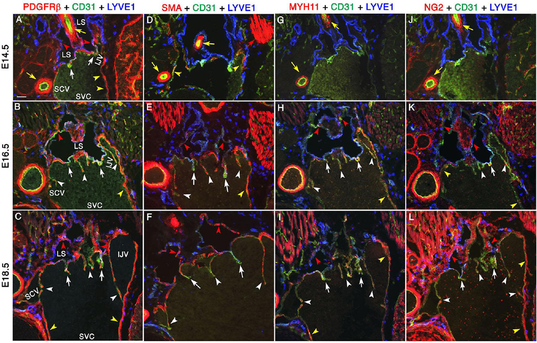
Figure 5. Mural cells are progressively recruited to lymph sacs, LVVs and VVs.
Mural cells are progressively recruited to lymph sacs, LVVs and VVs. LVV-forming region of E14.5, E16.5 and E18.5 embryos were analyzed using mural cell markers. PDGFRβ (A-C) is a pan-mural cell marker. SMA (D-F) and MYH11 (G-I) are vascular smooth muscle cell markers. NG2 is a pericyte marker (J-L). At E14.5, PDGFRβ+ cells are is seen surrounding the lymph sacs (A, red arrowheads) and within LVVs (A, white arrows). Other mural cell markers are not seen in lymph sacs or LVVs at this stage (D, G, J). At subsequent developmental time points, downregulation of LEC marker LYVE1 in lymph sacs coincides with the expression of mature mural cell markers (red arrowheads). Scattered mural cells are also seen within LVVs (white arrows) and VVs (white arrowheads). Yellow arrows and yellow arrowheads point to the arterial and venous perivascular cells respectively. And, red arrows point to the muscles.
Statistics: n= 3 for each developmental stage.
Abbreviations: LS, lymph sac; IJV, internal jugular vein; EJV, external jugular vein; SCV, subclavian vein; SVC, superior vena cava.
Scale bars: 50 µm

