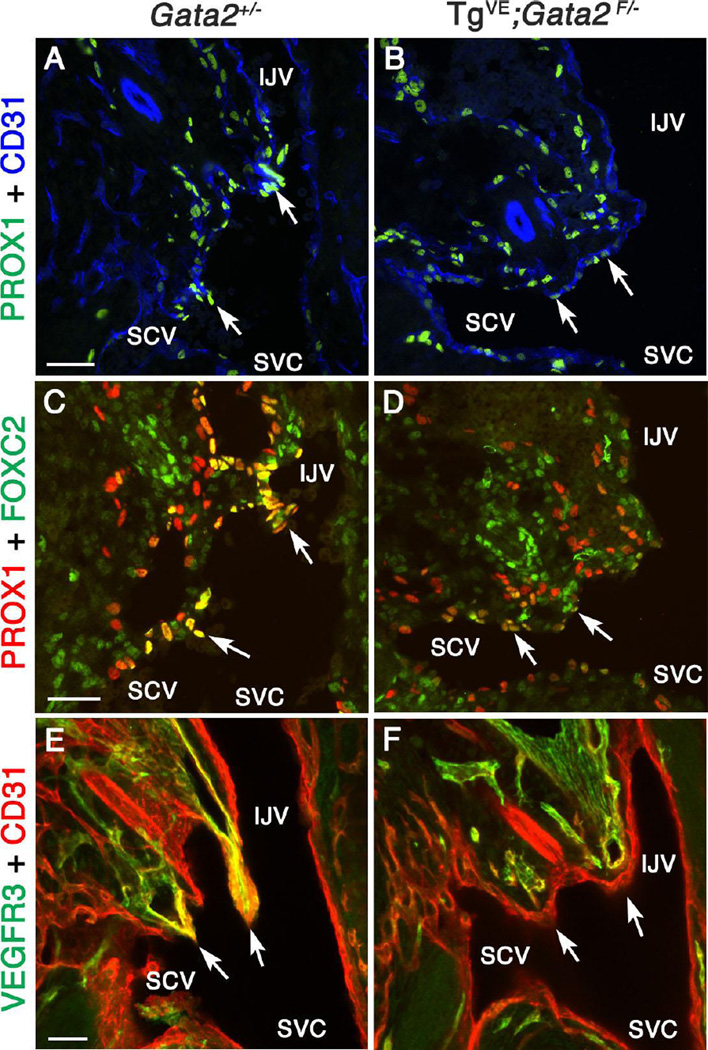
Figure 9. GATA2 is necessary for the proper differentiation of LVV-ECs.
(A-F) E13.5 Gata2+/− or TgVE;Gata2f/− (in which Gata2 is conditionally deleted from all endothelial cells using CreERT2 after tamoxifen injection at E10.0) embryos were analyzed using the indicated markers. A-D are 12 µm frontal cryosections. PROX1 (A, C arrows) and FOXC2 (C, arrows) are strongly expressed in LVV-ECs of Gata2+/− embryos. In contrast, PROX1 (B, D arrows) and FOXC2 (D, arrows) are weakly expressed in the LVV-forming region of TgVE;Gata2f/− embryos. E and F are projections of 100 µm thick frontal cryosections imaged by confocal microscopy. LVVs (E, arrows) seen in Gata2+/− are absent in TgVE;Gata2f/− embryos (F, arrows).
Statistics: n= 3.
Abbreviations: LS, lymph sac; IJV, internal jugular vein; SCV, subclavian vein; SVC, superior vena cava.
Scale bars: 50 µm.

