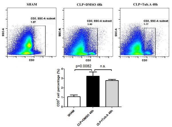
Figure 5. Tubastatin A treatment did not alter the percentages of T lymphocytes.

Representative plots T lymphocytes are shown on the right side of panel (CD3+). The percentages of T lymphocytes were quantified and compared among groups. CLP: cecal ligation and puncture; Tub.A: Tubastatin A; SSC: side scatter.
