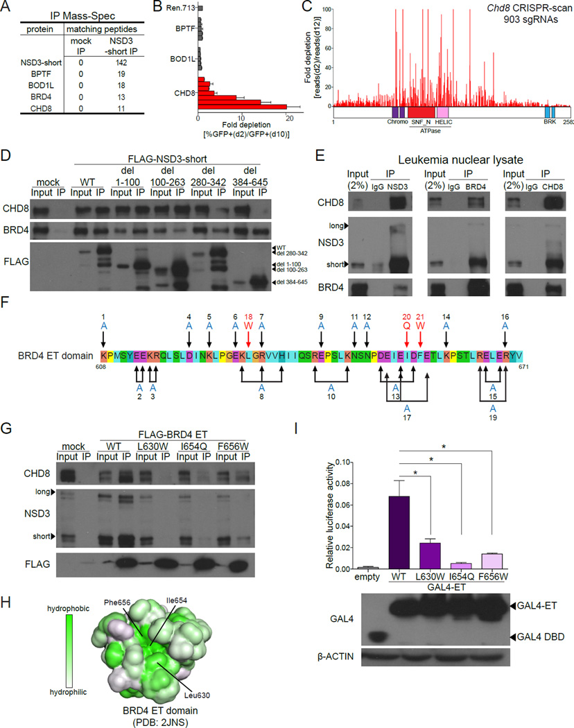
Figure 2. NSD3-short is an adaptor protein that links BRD4 to the CHD8 chromatin remodeling enzyme.
(A) Mass spectrometry analysis of proteins identified using anti-FLAG IP performed with nuclear lysates prepared from HEK293T cells transfected with FLAG-NSD3-short or empty vector (for mock IP). The list was ranked by the total number of matched peptides recovered. Complete results are provided in Table S1. (B) Competition-based assay in RN2 cells evaluating the effect of LMN shRNAs targeting the indicated proteins. Each bar represents the average fold-decrease in the percentage of GFP+ cells over 8 days for individual shRNAs. n=3. (C) CRISPR-scan of Chd8 with all possible sgRNAs. Deep sequencing based measurement of the impact of 907 Chd8 sgRNAs on the proliferation of Cas9-expressing RN2 cells. The location of each sgRNA relative to the CHD8 protein is indicated along the x axis. Shown is a representative experiment of two biological replicates. (D) IP-Western blotting performed with anti-FLAG antibodies and nuclear lysates prepared from RN2 cells stably expressing the indicated FLAG-NSD3 constructs or empty vector. (E) Endogenous IP-Western blotting performed with the indicated antibodies and nuclear lysates prepared from NOMO-1 cells. (F) The amino acid sequence of the human BRD4 ET domain indicating the surface residues that were subjected to mutagenesis. Combinations of mutations were used in some cases in an attempt to disrupt specific clusters of charged residues. (G) IP of the indicated FLAG-BRD4 ET domains expressed transiently in HEK293T cells followed by Western blotting with the indicated antibodies. (H) The molecular surface of the BRD4 ET domain (PDB: 2JNS) with hydrophobicity indicated in green (Lin et al., 2008). (I) (top) Luciferase reporter assay evaluating the activation function of the indicated GAL4-ET domain fusions on a minimal plasmid-based reporter harboring GAL4 recognition sequences. HEK293T cells were co-transfected with p9xGAL4-UAS-luciferase (firefly) reporter and the indicated GAL4 fusion expression plasmids expressing Renilla luciferase from a constitutive promoter. Plots indicate firefly luciferase activity normalized to the Renilla luciferase control. n=3. *p<0.05, two-tailed Student’s t-test. (bottom) Western blotting analysis of HEK293T cells transfected with the indicated plasmids shown in the top panel. All error bars represent SEM and all IP-Western and Western blotting experiments shown are a representative experiment of at least three independent biological replicates. See also Figure S2.

