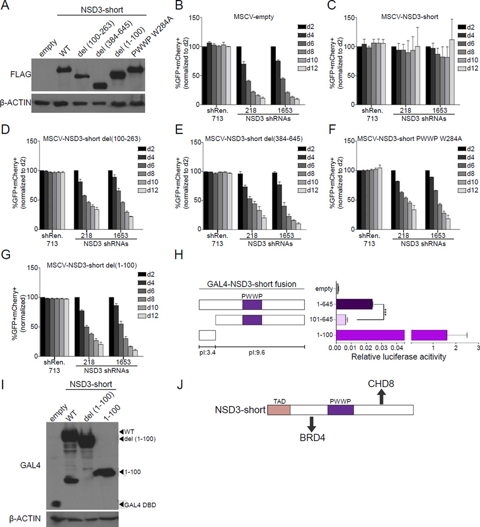
Figure 3. NSD3-short utilizes four distinct interaction surfaces to sustain AML cell proliferation.
(A) Western blotting analysis of whole cell lysates prepared from RN2 cells transduced with the indicated PIG retroviral expression constructs. A representative experiment of three independent biological replicates is shown. (B–G) Competition-based assay tracking the abundance of GFP+mCherry+ cells during culturing of transduced RN2 cells. GFP is linked to the indicated cDNA and mCherry is linked to the indicated LMN shRNA. Plotted is the average of three independent biological replicates, normalized to d2. (H) Luciferase reporter assay with the indicated GAL4 fusions, performed as described in Figure 2I. n=3. ***p<0.001, two-tailed Student’s t-test. (I) Western blotting analysis of HEK293T cells transfected with the indicated plasmids. A representative experiment of three biological replicates is shown. (J) Diagram of the functionally important surfaces of NSD3-short. All error bars in this figure represent SEM. See also Figure S3.

