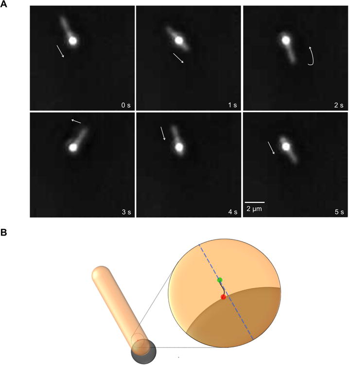
Figure 1.

Movement of F. johnsoniae on a stationary polystyrene bead. (A) Images at 1 s time intervals from Movie S1, taken using a phase contrast microscope with a bright-phase objective (Nikon Plan 40× BM), showing a rod-shaped F. johnsoniae cell (slightly out of focus) moving over a spherical bead (0.5 μm). Time is shown at the bottom right of each frame and a scale bar is shown at bottom left of the last frame. Arrows depict direction of motion of the leading pole (0-3 s), which later becomes the lagging pole (4 s). (B) Diagrammatic representation of a cell moving over a stationary bead, with a magnified view in the inset. In the magnified view, a SprB filament (black), which is underneath the cell, is bound to the bead at an attachment site (red dot) that remains fixed. The SprB filament is loaded on a tread guided by a track (blue dotted line), and the SprB filament-to-tread attachment site (green dot) is in motion relative to the bead.
