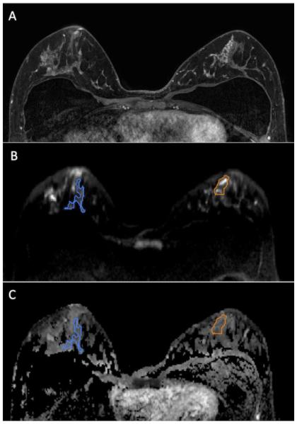
Figure 1.
47 year-old woman with segmental non mass enhancement in the left breast spanning 40 mm on MRI (A), biopsy-proven low risk DCIS (Van Nuys Pathological Classification 1). Diffusion weighted images show a contrast-to-noise ratio of 3.0 (B) with ADC value of 1.46 × 10⁻³ mm2/s and normalized ADC value of 0.80 (C). Note: Normal breast tissue ROI is shown in blue while the ROI for the lesion is shown in orange.

