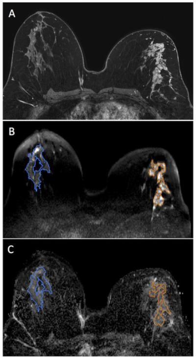
Figure 2.
46 year-old woman with segmental non mass enhancement in the left breast spanning 105 mm on MRI (A). Biopsy yielded higher risk DCIS (Van Nuys Pathological Classification 2). Diffuse weighted images show a contrast-to-noise ratio of 2.29 (B) with ADC value of 1.63 × 10⁻³ mm2/s and normalized ADC value of 1.13 (C). Note: Normal breast tissue ROI is shown in blue while the ROI for the lesion is shown in orange.

