Figure 4.
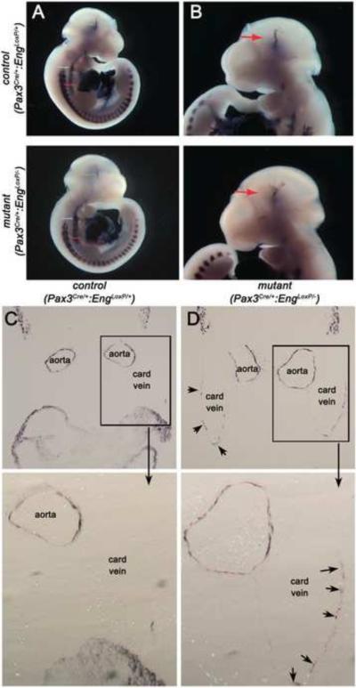
Loss of endoglin expression in Pax3Cre-expressing cells alters smooth muscle cell development and smooth muscle actin expression. (A, B) Whole mount vascular smooth muscle alpha actin (αSM-actin) staining of control Pax3Cre/+;EngLoxP/+ and mutant Pax3Cre/+;EngLoxP/− E10.5 embryos. Arrows point to (A) dilated dorsal aortae and (B) cranial vessels. (C, D) Sections showing αSM-actin positive cells in Pax3Cre/+;EngLoxP/+ (C) and mutant Pax3Cre/+;EngLoxP/− (D) E10.5 embryos. Distension of the paired aortae is apparent in the mutant, with ectopic SM-actin staining in the cardinal veins (arrows, card vein). Boxed areas are shown at higher magnification underneath.

