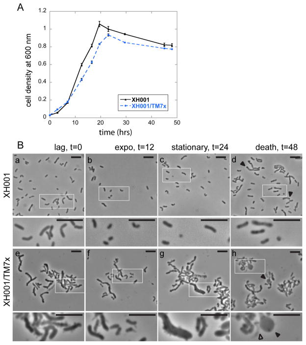
Figure 1.
Growth and morphology of XH001 monoculture and XH001/TM7x co-culture. (A) Mono (triangle, black solid line) and co-culture (circle, blue broken line) cell densities were determined by measuring the optical density at 600 nm (OD600). Each point represents the average of three independent cultures (error bars, SD). The time points were connected by straight line to guide the eye. (B, a–d) XH001 monoculture grown in microaerophilic conditions has short rod morphology from early to late growth phases. The lower images are a higher magnification of the boxed region in the upper image. During death phase (B, d), many of the cells had condensed dot structures within the cell body (arrow heads). (B, e–h) TM7x-associated XH001 have elongated and hyphal morphology from early to late growth phases. During exponential phase (B, f), many of the XH001 cells are short rods similar to XH001 alone. At death phase (B, h), many of the XH001 cells display large clubbed-ends (arrow heads), swollen cells, and cell lyses (open arrow head). During all growth phases, XH001 was heavily decorated with TM7x. All scale bars indicate 5 μm.

