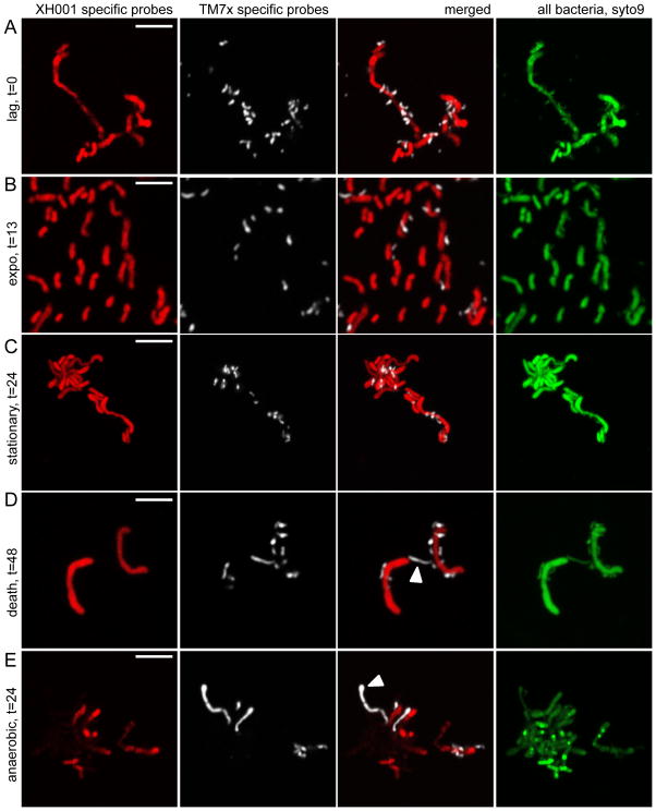
Figure 5.
Morphology of TM7x during growth. Co-culture grown under microaerophilic conditions was FISH stained using TM7x-specific (white, TM7567) and XH001-specific (red, M33910) DNA-probes that target the 16S rRNA gene (see Materials and Methods). Green is a universal DNA dye, syto9, which stains both XH001 and TM7x DNA and RNA. XH001 monoculture staining is shown in Figure S3. At time points 0 (A), 24 (C) and 48 (D) hours, XH001 had long and hyphal morphology. At 13 hours (B), many of the XH001 cells were short rods. TM7x also assumed different morphologies. At time points 0–24 (A–C) hours, TM7x appeared as small cocci or short rods. At 48 hours (D), the elongated form of TM7x was observed more frequently (white arrow head). (E) Interestingly, TM7x also assumed an elongated morphology when the co-culture was incubated under anaerobic conditions (white arrowhead). Long TM7x can be observed throughout all time points of the growth but only t = 24 hours is shown in E. All scale bars indicate 5 μm.

