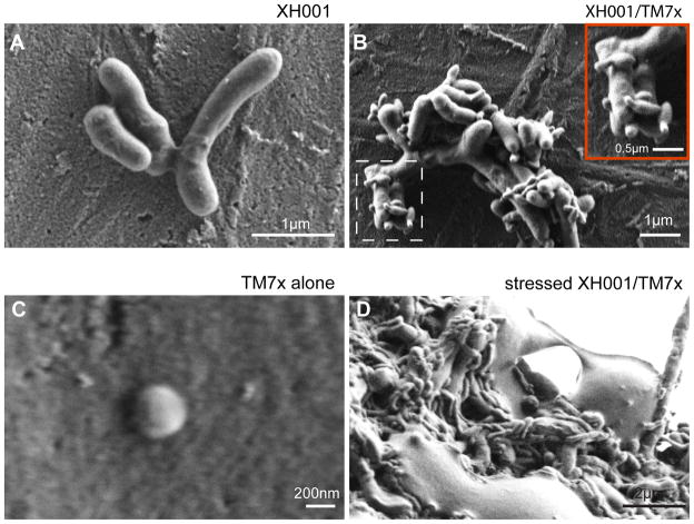
Figure 7.
Morphology of XH001 and TM7x under SEM. (A) XH001 monoculture grown under microaerophilic conditions shows short rod morphology with smooth surfaces. (B) Co-culture grown under microaerophilic conditions reveals elongated and branched XH001 cells decorated with smaller cells, presumably TM7x. Inset shows a zoomed-in image of the smaller TM7x (box with dashed line) that seems to be budding. (C) Isolated TM7x using 0.22-micron filter shows cocci TM7x with diameter of ~200 nm. (D) Co-culture grown under anaerobic conditions shows elongated XH001 decorated with many elongated TM7x.

