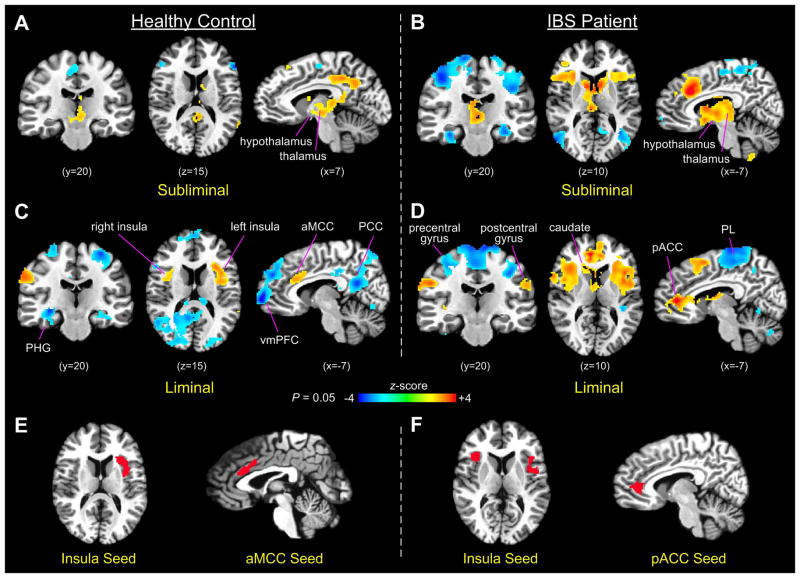
Figure 2.
(A–D) Brain activation in controls and IBS patients during subliminal and liminal rectal distensions (PL, parietal lobule; PHG, parahippocampal gyrus). (E) Seed regions defined in the anterior insula (left) and aMCC (right) based on rectal-distension-induced activation in the control group. (F) Seed regions defined similarly in the bilateral anterior insula (left) and pACC (right) in the IBS patient group.

