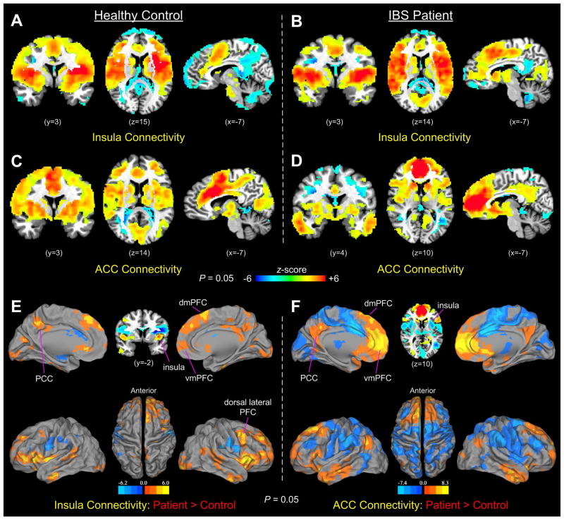
Figure 4.
(A–B) Functional connectivity of the insula seeds in the control and IBS groups during liminal stimulation. (C–D) Functional connectivity of the aMCC (in controls) and pACC (in IBS patients) seeds during liminal stimulation. (E–F) Group comparisons of insular and cingulate functional connectivity between controls and IBS patients. In both comparisons, IBS patients showed significantly increased functional connections in the dmPFC, vmPFC, dorsal lateral PFC, and PCC, compared with controls.

