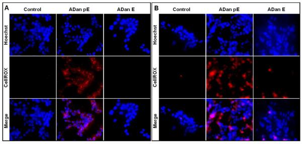
Figure 8. Oxidative stress in cells challenged with ADan.
SH-SY5Y cells were challenged with ADan pE/E (50 μM) for 2h (A) or 4h (B) followed by staining with the CellROX Deep Red fluorogenic probe. In (A) and (B) Top panel: Hoechst nuclear stain shown in blue; Central panel: oxidized CellROX reagent highlighted in red; Bottom panel: merged images. Magnification = X20 in all cases.

