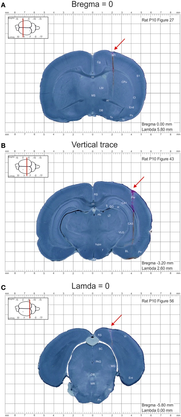
Figure 1.

Example images of the coronal sections from the postnatal day P10 rat brain from the atlas of the developing rat brain in stereotaxic coordinates. (A–C) Coronal sections at anterioposterior zero distances from the bregma (A) and lambda (C), and a section illustrating the trace through the whole brain (B). DiI-labeled penetrations are indicated by red arrows.
