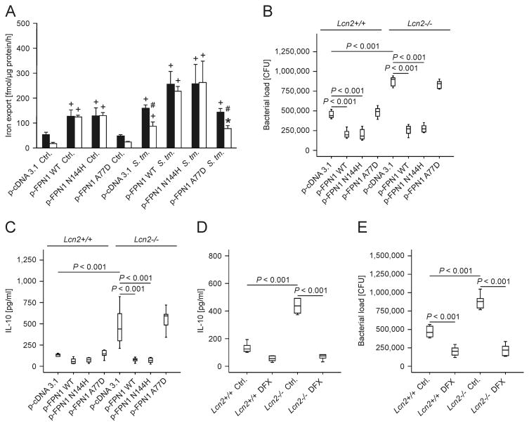
Figure 4. Forced expression of FPN1 or iron chelation by DFX rescues the phenotype of Lcn2-/- peritoneal macrophages.
(A) Wt (closed bars) and Lcn2-/- peritoneal macrophages (open bars) were transiently transfected with empty plasmid (p-cDNA3.1) or expression constructs containing the indicated human FPN1 variants. 59Fe transport studies were used to determine macrophage iron release following treatment with diluent (Ctrl.) or infection with wt Salmonella Typhimurium (S. Tm.). Values are depicted as means ± S.E.M. and statistically significant differences as calculated by ANOVA are indicated (n = 4 independent experiments). # P value < 0.01 for the comparison of wt and Lcn2-/- peritoneal macrophages. + P value < 0.001 as compared to the control of the respective genotype. * P value = 0.003 as compared to the control of the respective genotype. (B, C) The bacterial load (B) in macrophages was quantified by plating of cell lysates on LB agar and the accumulation of IL-10 (C) in cell culture supernatants was measured by an ELISA kit. Values are depicted as as means ± S.E.M. lower quartile, median and upper quartile (boxes) with minimum and maximum ranges and statistically significant differences are indicated (n = 6 independent experiments). (D, E) The bacterial load and IL-10 production was quantified following treatment with 50 μM DFX (n = 6 independent experiments). Bars show lower quartile, median and upper quartile (boxes) with minimum and maximum ranges and statistical significant differences as determined by ANOVA are indicated.

