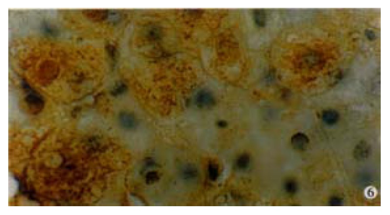
Figure 6.

Immunohistochemical preperation stained by specific HGV McAb for HGVNS5 in a patient with single HG infection showing brown-yellow granules presented mostly in cytoplasm of hepatocytes, and partially in the nuclei. DAB staining, hematoxylin staining, BA staining. (oil) × 1000
