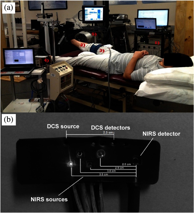
Fig. 1.
(a) Experimental setup: (1) diffuse correlation spectroscopy (DCS)/near-infrared spectroscopy (NIRS) hybrid equipment received analog position input from the dynamometer to gate data collection during exercise. (2) Optical probe [depicted uncovered in (b)] sited over medial gastrocnemius at the location of the muscle belly. (3) Occlusion cuff secured above the thigh for inducing venous and arterial occlusions (VO and AO) for calibration. (4) The strain gauge and control computer run independently and concurrently during both calibration and exercise protocols. (5) The dynamometer was used to control exercise workload and frequency via setting 30% maximal voluntary isometric contraction (MVIC) resistance and passive return to rest. A visual metronome was used to regulate subject timing.

