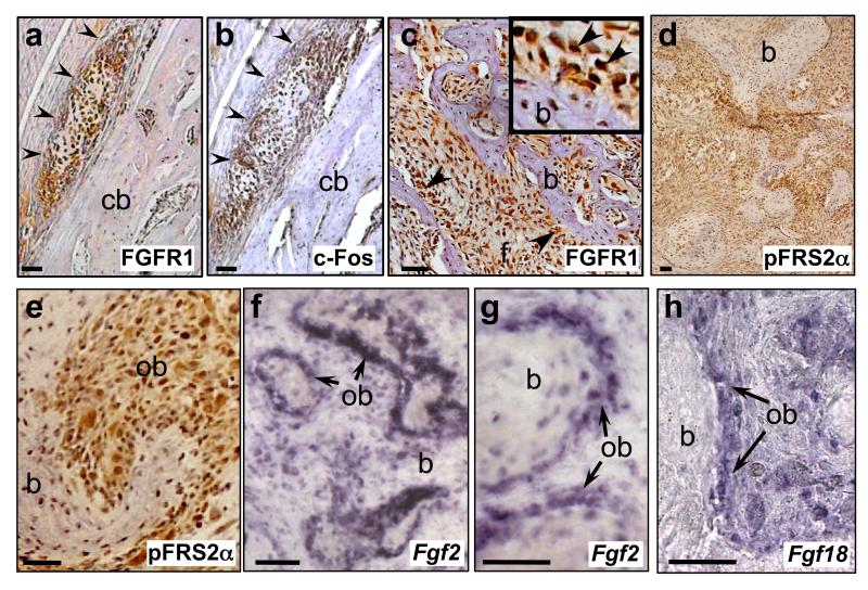
Figure 4.
Expression of FGF/FGFR signalling components in c-Fos-transgenic mouse osteosarcomas and human tissue microarrays (TMA). (a-e) Immunohistochemical analysis of FGFR1, c-Fos and pFRS2α proteins as indicated, in early osteosarcoma lesions (a,b, arrowheads) arising adjacent to a femoral cortical bone surface (cb), as well as in late-stage c-Fos transgenic osteosarcomas (c-e) (see also Supplementary Figures 4a-c). FGFR1 (c) and pFRS2α (e) immunoreactive cells, including nuclear localisation, are observed in transformed osteoblasts (ob) lining neoplastic bone (b) (arrows, inset in (c)) as well as in fibrous areas (f). RNA in situ hybridisation for Fgf2 (f,g) and Fgf18 (h) expression in late-stage c-Fos transgenic tumours, showing high levels of FGF ligands in transformed osteoblasts (ob).

