Figure 7.
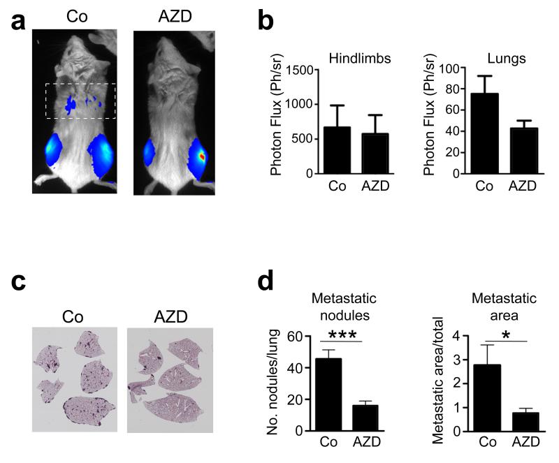
Effects of in vivo pharmacological inhibition of FGFR signalling on lung metastasis. (a) Representative bioluminescence images of Rag2-/-:IL2Rγ-/- immunocompromised mice (n=7/group) 14d after intratibial injection of wild-type P1.15 osteosarcoma cells followed by daily administration of either vehicle (Co) or the FGFR inhibitor AZD4547 (AZD). The dotted rectangle identifies the bioluminescence imaging area of the thoracic cavity/lungs. (b) Quantification of bioluminescence in the hindlimbs and lungs. (c) Representative photomicrographs of H&E-stained histological sections of lungs, showing metastatic nodules formed in mice 14d after intratibial injection of wild-type P1.15 osteosarcoma cells followed by daily administration of either vehicle (Co) or the FGFR inhibitor AZD4547 (AZD). (d) Quantification of the number of metastatic nodules, and the percent metastatic area per lung formed in the lungs of animals injected with P1.15 cells and treated with AZD4547 as above. All data represent the mean ± SD of triplicate samples. *p < 0.05, ***p < 0.001 vs respective controls.

